
Project · Results
Results
Data, figures, and interpretation, with what is proof-of-concept vs. preliminary stated clearly.
Twenty-one figures, and what each one proves. Every experimental figure from the season, in the order the work happened, with what it shows and what it supports. Nothing is claimed beyond what its figure carries.
Abstract
We set out to express fatty acid amide hydrolase from Faecalibacterium prausnitzii in E. coli Nissle 1917, and to find out where in the cell the enzyme has to sit in order to reach oleoylethanolamide in the gut lumen.
Across the season we designed a six-construct localisation panel, amplified as twelve PCR reactions, assembled constructs by in vivo assembly cloning into pEM106.2, and verified them by restriction digest and Sanger sequencing. Four constructs are complete and sequence-verified. Three of them express protein of the expected mass on induction. One, the YebF secretion variant, has been transformed into our target chassis and banked.
We have not yet measured enzyme activity. The figures below report what we built and what we observed, and the conclusions are limited accordingly.
Main results
- Four constructs sequence-verified — 6xHis-GFP, 6xHis-WT-FAAH, 6xHis-ΔTm-FAAH and 6xHis-YebF-ΔTm-FAAH.
- Expression confirmed by SDS-PAGE for GFP, WT-FAAH and ΔTm-FAAH in BL21(DE3), with induced bands absent from their own uninduced controls.
- A single-nucleotide insertion removed after eight weeks and three iterations, by moving the ligation out of the cell.
- Candidate probiotic strain built and banked — YebF-ΔTm-FAAH in EcN, five glycerol stocks at −80 °C.
Construct design and cloning strategy
We assemble into the pEM106.2 backbone by in vivo assembly cloning, replacing its YebF-hIL10 cassette with our insert. Backbone and insert are amplified separately with primers carrying 15–20 bp overlapping tails, the parental template is destroyed with DpnI, and the two linear fragments are co-transformed so the cell’s own recombination machinery joins them.
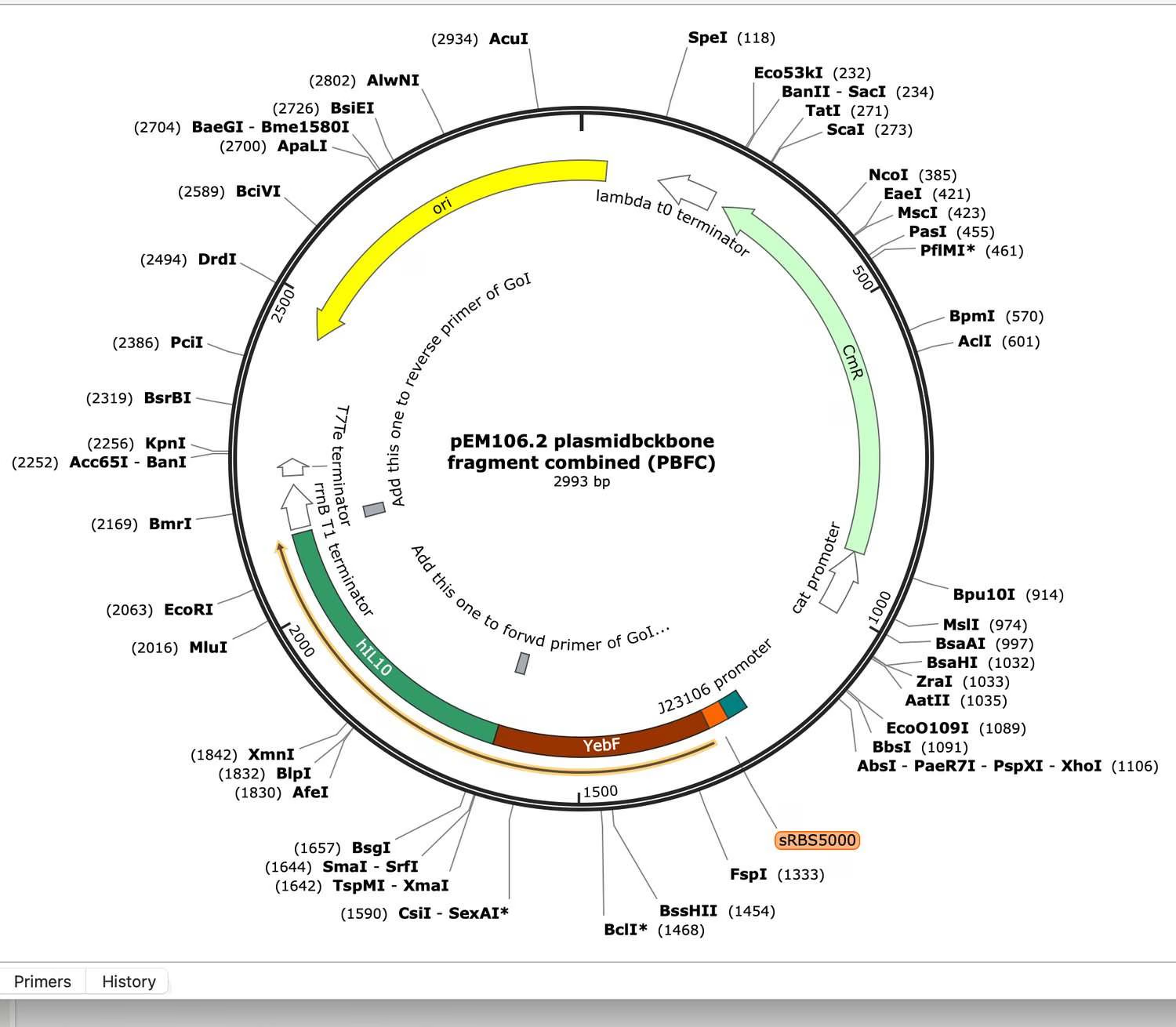
What this shows. The vector we clone into, with the restriction sites we mapped in order to choose screening enzymes, and the YebF-hIL10 cassette that our inserts replace.
Conclusion. The backbone supplies an inducible promoter, chloramphenicol selection and a secretion carrier already in position, which can be kept as the YebF variant or swapped for a membrane display tag.
The localisation panel
| Variant | Strategy | What it tests |
|---|---|---|
| 6xHis-WT-FAAH | Keeps its transmembrane sequence | Membrane-associated reference |
| 6xHis-ΔTm-FAAH | Transmembrane domain deleted | Does losing the anchor free it |
| 6xHis-YebF-ΔTm-FAAH | YebF secretion carrier | Can it be exported |
| 6xHis-OmpX-ΔTm-FAAH | OmpX signal peptide | Outer membrane display |
| 6xHis-OmpA-ΔTm-FAAH | OmpA signal peptide | Display, second route |
| 6xHis-GFP | Fluorescent expression reporter | Expression and backbone control |
| Catalytic mutant | Dead active site | Is the activity really FAAH |
Six of these carry designed primers. The catalytic mutant does not; it was specified in the panel but never reached primer design, which is why it appears as Not built in the status table.
Why two of these are controls
6xHis-GFP reports inducible expression in the backbone independently of whether the enzyme works. The catalytic mutant allows any activity we measure to be attributed to FAAH rather than to a host enzyme. A localisation panel without both is not interpretable.Amplifying the constructs
Most reactions amplified under the standard protocol. Reaction #7, the backbone fragment for one of the FAAH variants, did not.
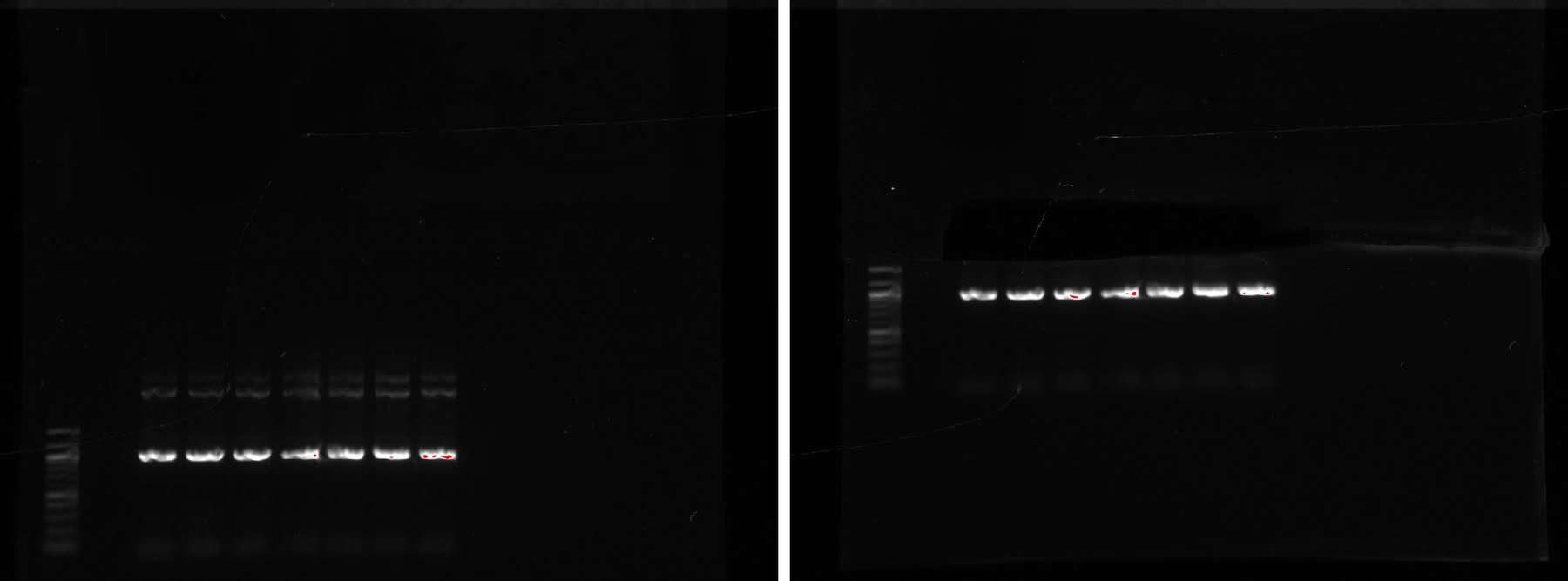
What this shows. Product is present at every temperature in the gradient, so the reaction works, but each lane carries additional bands above the expected product.
Conclusion. The failure was not a lack of amplification but a lack of specificity. Annealing temperature alone did not resolve it.
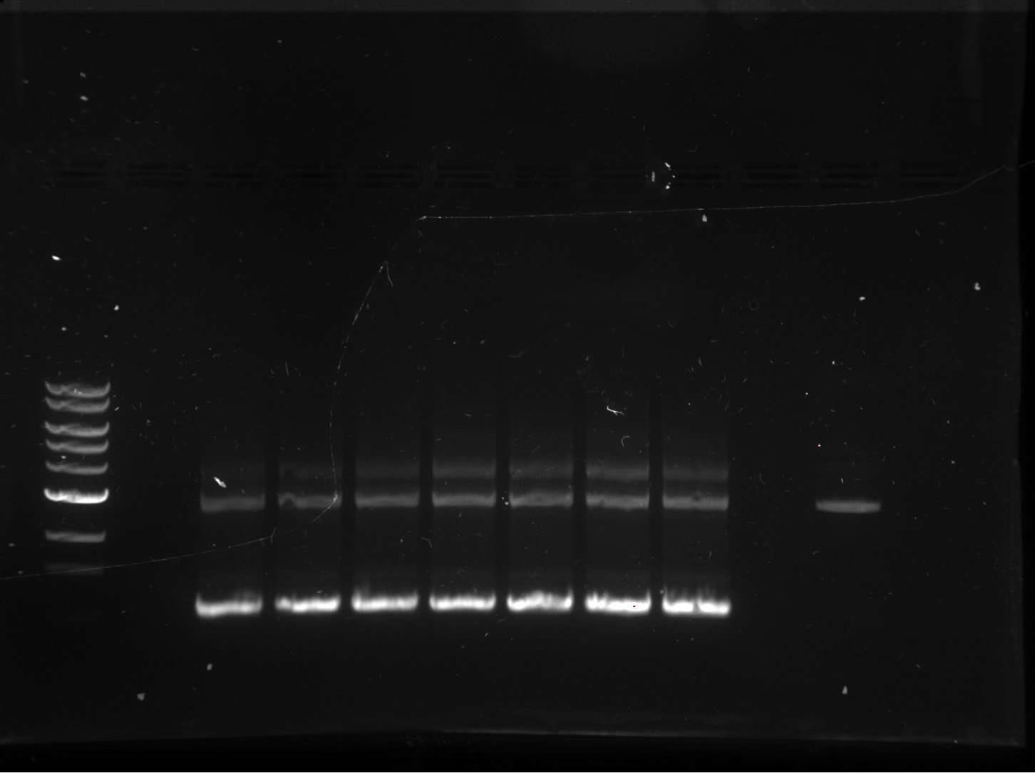
What this shows. Raising the cycle count from 17 to 20 gives a stronger product sitting near the expected size when read against the 10 kb and 3 kb plasmid references rather than against the ladder alone.
Conclusion. Running the parent plasmid as a size reference in the same gel was more informative than the ladder for a fragment of this size, and we adopted it as routine.
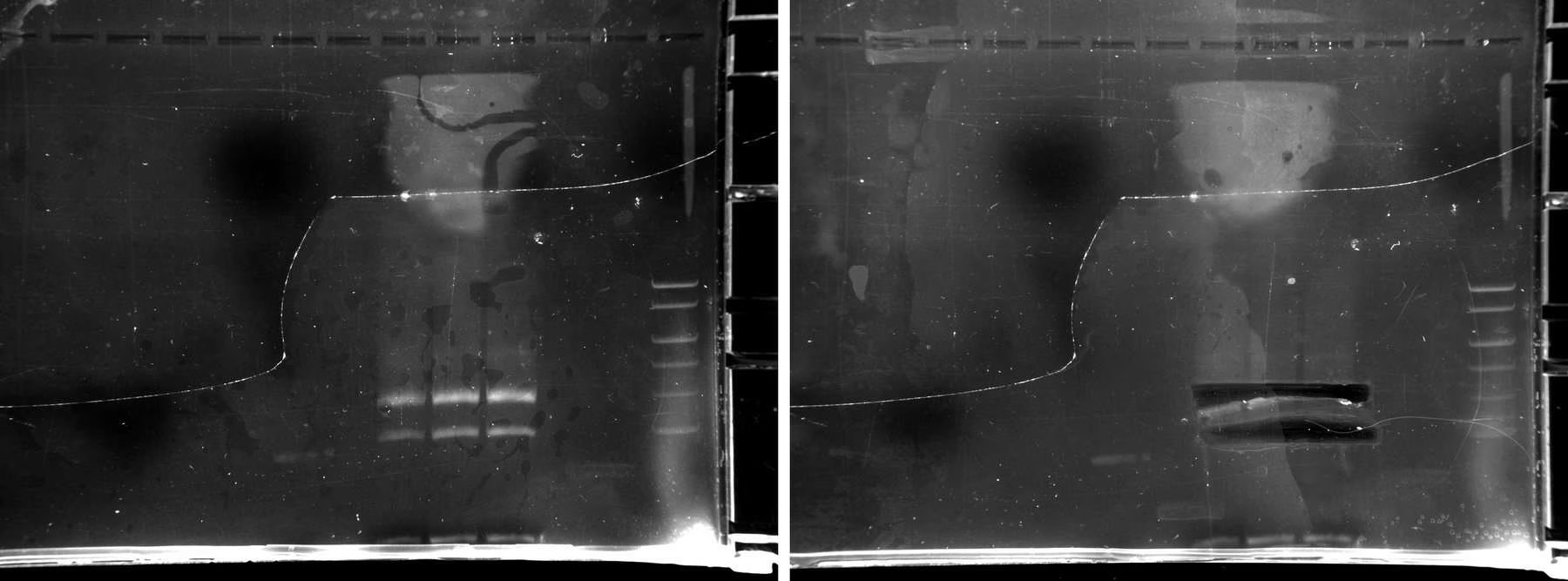
What this shows. At 65 °C the two upper bands separate enough to be cut out individually. Gel extraction recovered 8 ng/µL from the upper band and 13 ng/µL from the lower band, roughly 30 µL each.
Conclusion. Yield was low but sufficient to transform. The upper band went on to produce the clones carrying the correct insert, so this separation is the step that rescued the construct.
Ampicillin produced satellite colonies
Our first transformations were plated on 100 µg/mL ampicillin. Colonies appeared and looked normal, but none established in liquid culture.
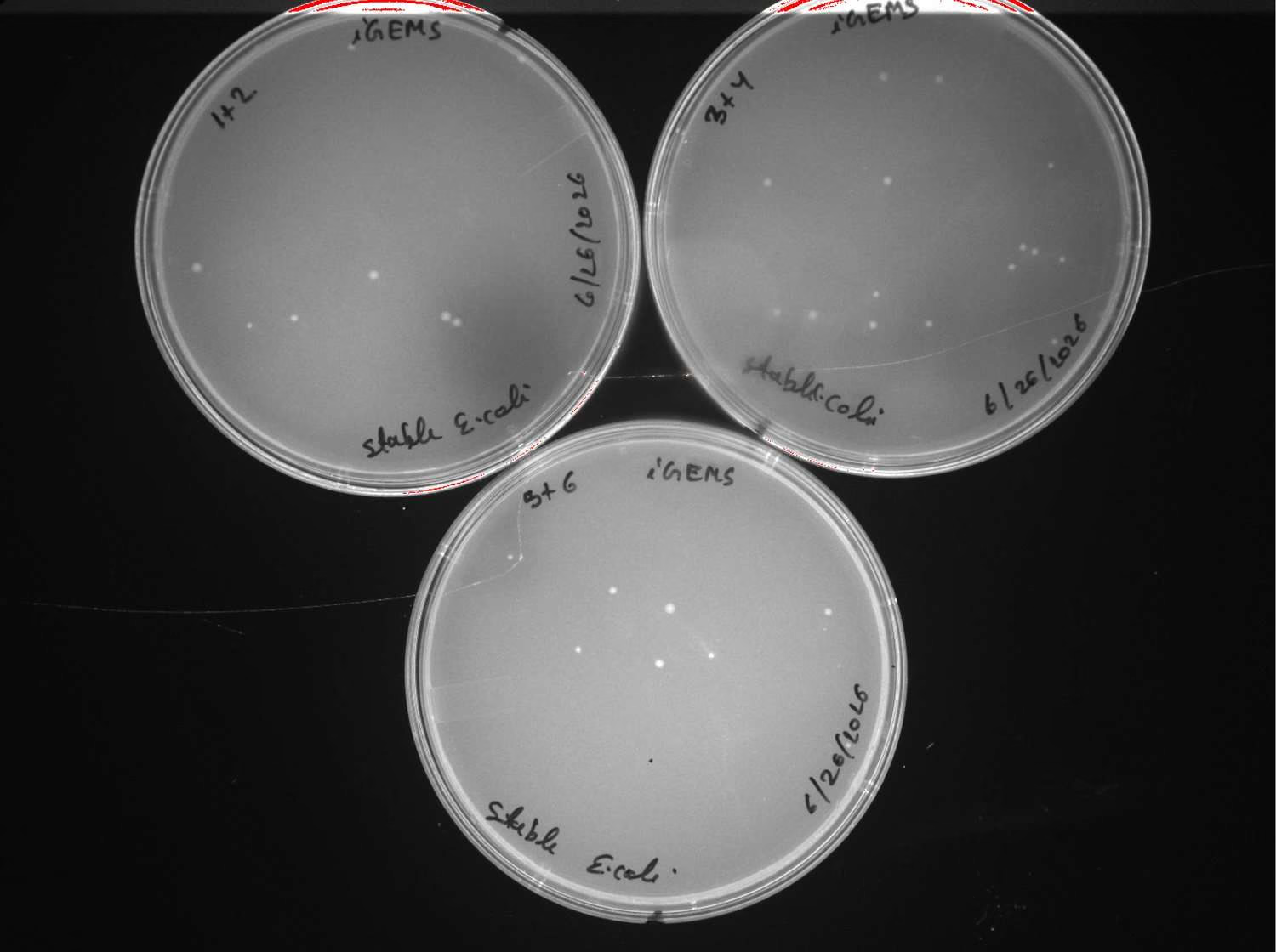
What this shows. Colonies are present but few, and clustered rather than evenly distributed. None grew when inoculated into LB with ampicillin.
Conclusion. These are satellite colonies: cells growing where a genuine transformant has already degraded the surrounding ampicillin through secreted beta-lactamase. They are not resistant themselves and are indistinguishable from real transformants on a plate.
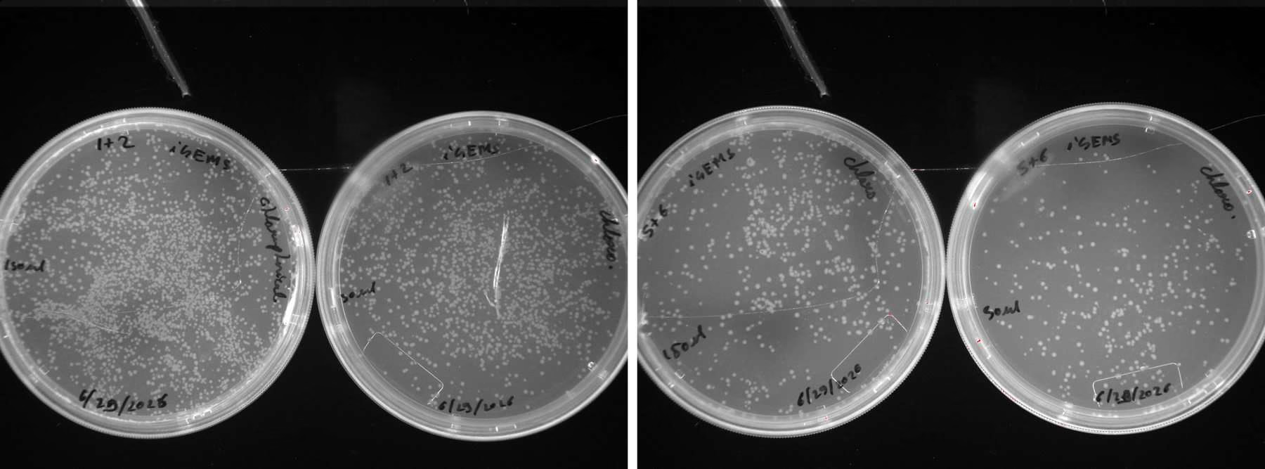
What this shows. Growth is far heavier and evenly distributed, and these colonies established normally in liquid culture.
Conclusion. The transformations had been working all along; the selection was reporting them incorrectly. Chloramphenicol was used for the rest of the season and the problem never recurred.
Screening by restriction digest
Before committing clones to sequencing we screened miniprep DNA by restriction digest, choosing enzyme pairs that give a visibly different banding pattern for correct insert versus empty vector.
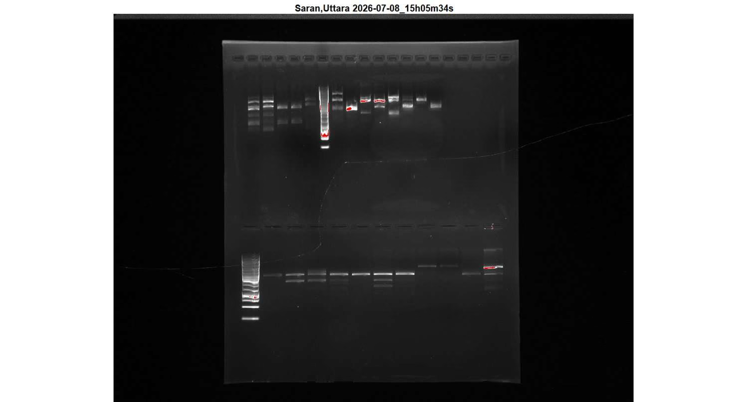
What this shows. Digest products across the screened minipreps, with banding patterns comparable against the predicted pattern for a correct construct.
Conclusion. The digest narrowed which clones were worth sequencing, but resolution on our gels was not good enough to call a construct correct on this evidence alone. Sequencing remained the deciding test throughout.
Sequence verification
Sanger sequencing across the insert junctions. These results determined which clones went forward, and are reported without figures because the primary data are reads rather than images.
Both rounds used the same clone numbering. The second round re-sequenced clones from the first with a different primer, which is why clones in the same number ranges are reported negative in Table 1 and confirmed in Table 2.
Table 1 · First round, submitted 17 July, returned 20 July
| Clones | Intended construct | Outcome |
|---|---|---|
| #1 – #5 | GFP | All negative or unrecorded read (#5) |
| #6 – #10 | WT FAAH | All negative |
| #11 – #15 | reaction 5+6 | All negative |
| #16, 17, 18, 20 | ΔTm-FAAH, lower band | Parent plasmid |
| #19, 21, 22, 23 | ΔTm-FAAH, upper band | Insert present, carries an extra 'A' |
| pEM106 | Positive control | Correct |
Twenty-four clones (K1 to K24) went out on this submission, together with the pEM106 control and the DF and DC samples.
Conclusion. The upper-band clones from Figure 4 carry the ΔTm-FAAH insert in frame with the parent plasmid, but with one additional adenine at the junction. The lower band was parent plasmid, which confirms that separating the two bands was necessary.
Table 2 · Second round, submitted 29 July, returned 31 July
| Clone | Construct | Outcome |
|---|---|---|
| #2 | GFP | Confirmed |
| #7, #8 | WT FAAH | Confirmed, #7 taken forward |
| #11, #12 | ΔTm-FAAH | Confirmed, #11 taken forward |
| #13, #14 | GFP, both termini clean | Confirmed, #13 taken forward |
Sixteen clones went out on this submission: K1 to K15 and K24, re-sequenced with a different primer. The four rows above are the clones that came back clean.
Adding the 6xHis tag
An N-terminal 6xHis tag was added to every construct so expression could be detected by Western blot and the protein purified later. The tag was introduced by whole-plasmid PCR with a shared reverse primer.
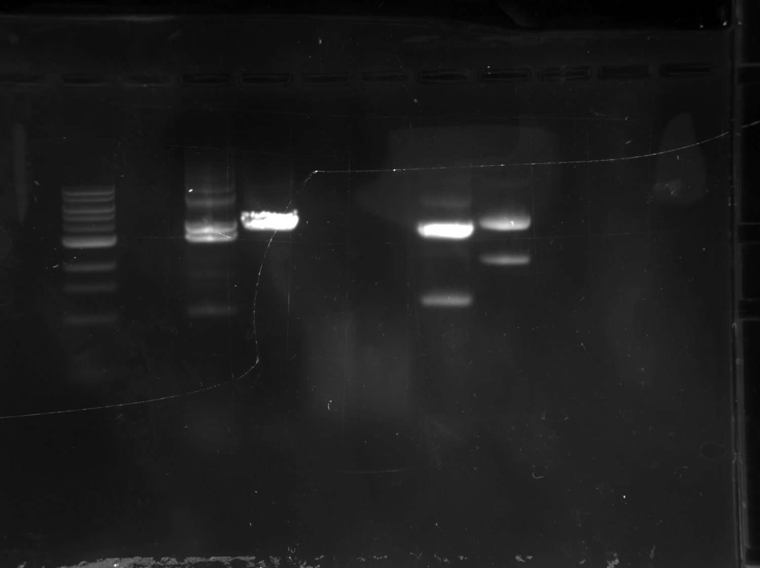
What this shows. The three 6xHis insertion reactions give clear product. The two 'A' removal lanes are empty.
Conclusion. Two reactions of the same design, run side by side under identical conditions, behaved completely differently. Tag insertion worked immediately; base deletion produced nothing, and became the problem that occupied the next eight weeks.
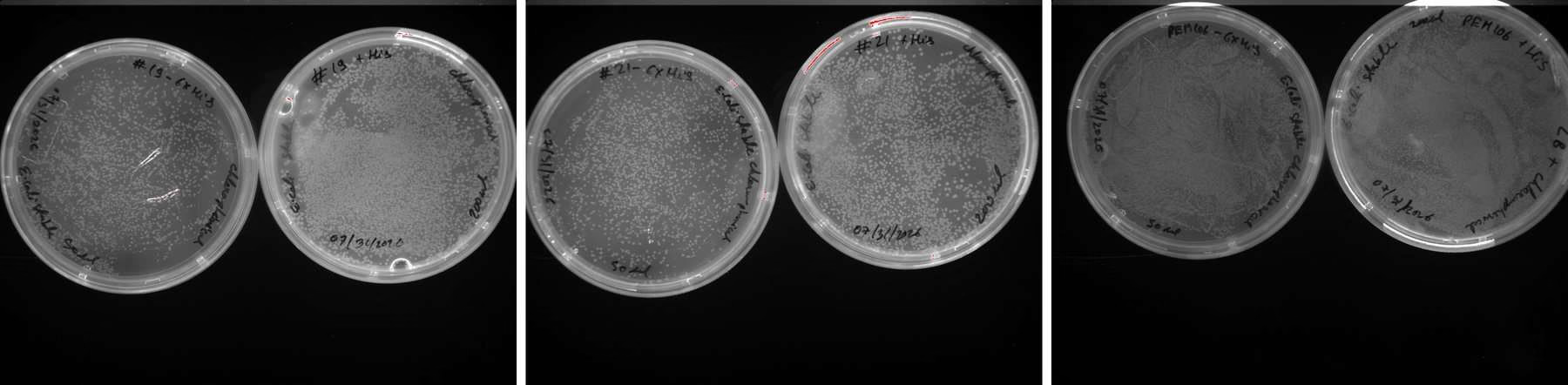
What this shows. Colonies recovered on all three constructs.
Conclusion. The tagged plasmids circularised and transformed successfully, which confirmed the tagging route before we committed the remaining clones to it.
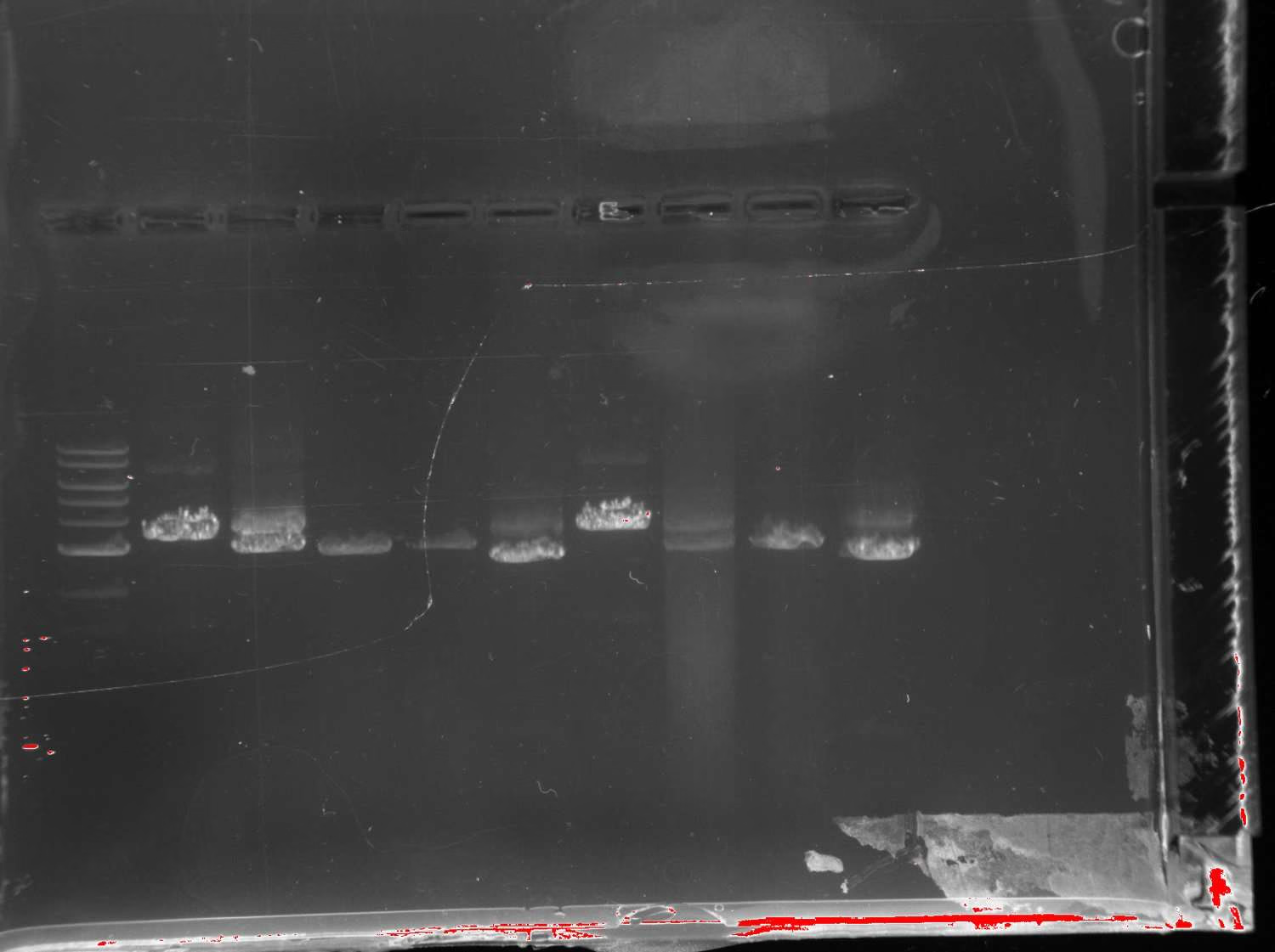
What this shows. Clean products near 3 kb for the tagging reactions at both annealing temperatures.
Conclusion. The tagging reaction is robust to annealing temperature across this range, so it did not require optimisation for each construct.
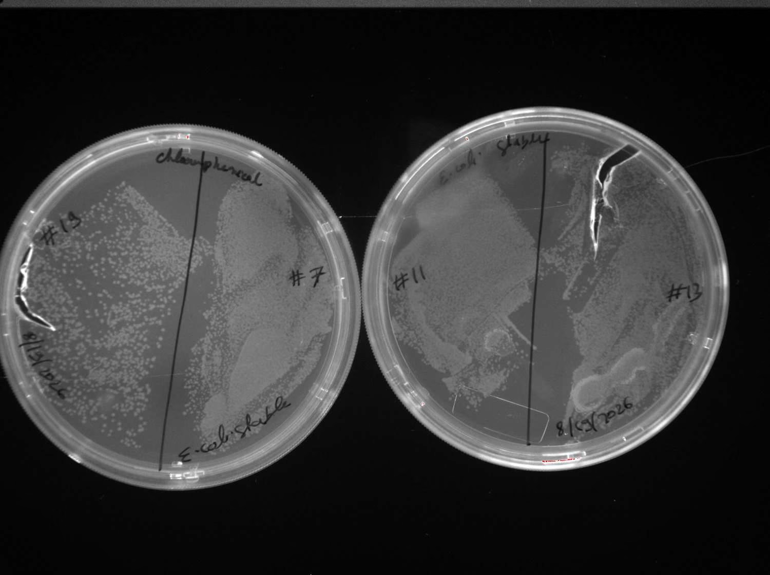
What this shows. Colonies on every sector, enough to pick three per construct.
Conclusion. Picking three rather than one mattered. Sequencing showed one #7 clone carried a spurious extra 'T', so a single pick would have been a coin flip. The same batch revealed a tube swap between #11_H2 and #13_H1, with both constructs correct but mislabelled, which is why we introduced a formal clone tracker.
Removing a single unwanted base
The ΔTm-FAAH junction carried one extra adenine, found in the first sequencing round returned on 20 July. Three rounds of whole-plasmid PCR relying on the cell to circularise the blunt product failed. The solution was to move the ligation out of the cell.
From the sequencing that found the extra base on 20 July to the sequencing that confirmed it gone on 15 September is eight weeks, followed by a final preparative confirmation on 21 to 23 September.
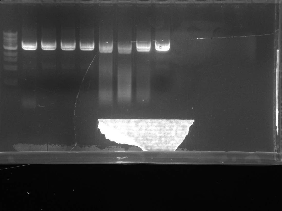
What this shows. Product across the gradient, so the amplification itself works at these temperatures.
Conclusion. Amplification was never the failing step. Sequencing of the resulting clones still showed the 'A' present, which pointed at what happens after the PCR rather than during it.
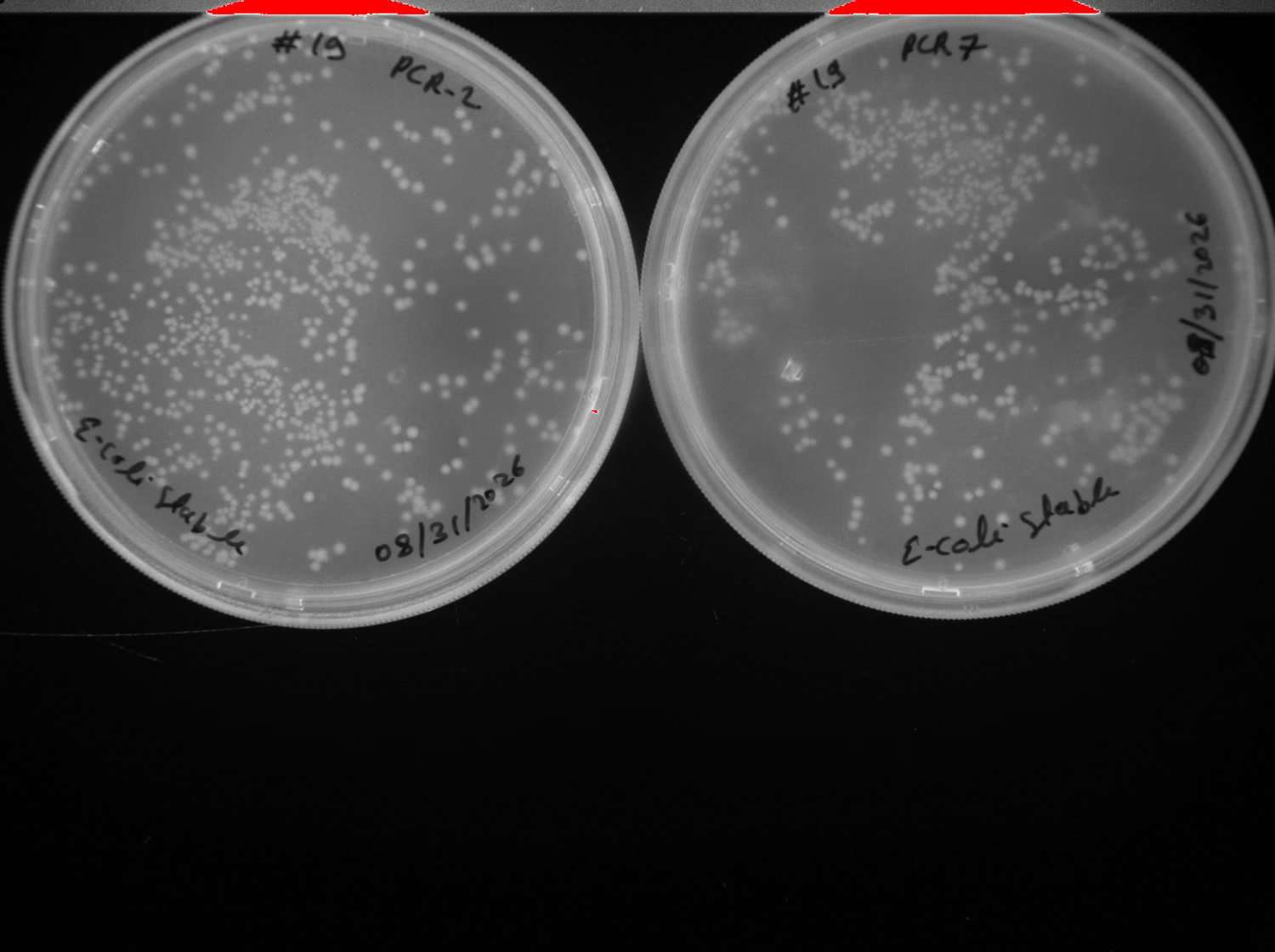
What this shows. Colonies recovered from the transformation.
Conclusion. Sequencing returned either unchanged parent plasmid or an ambiguous base call at the target position. Colonies were growing, but they were not carrying the edit.
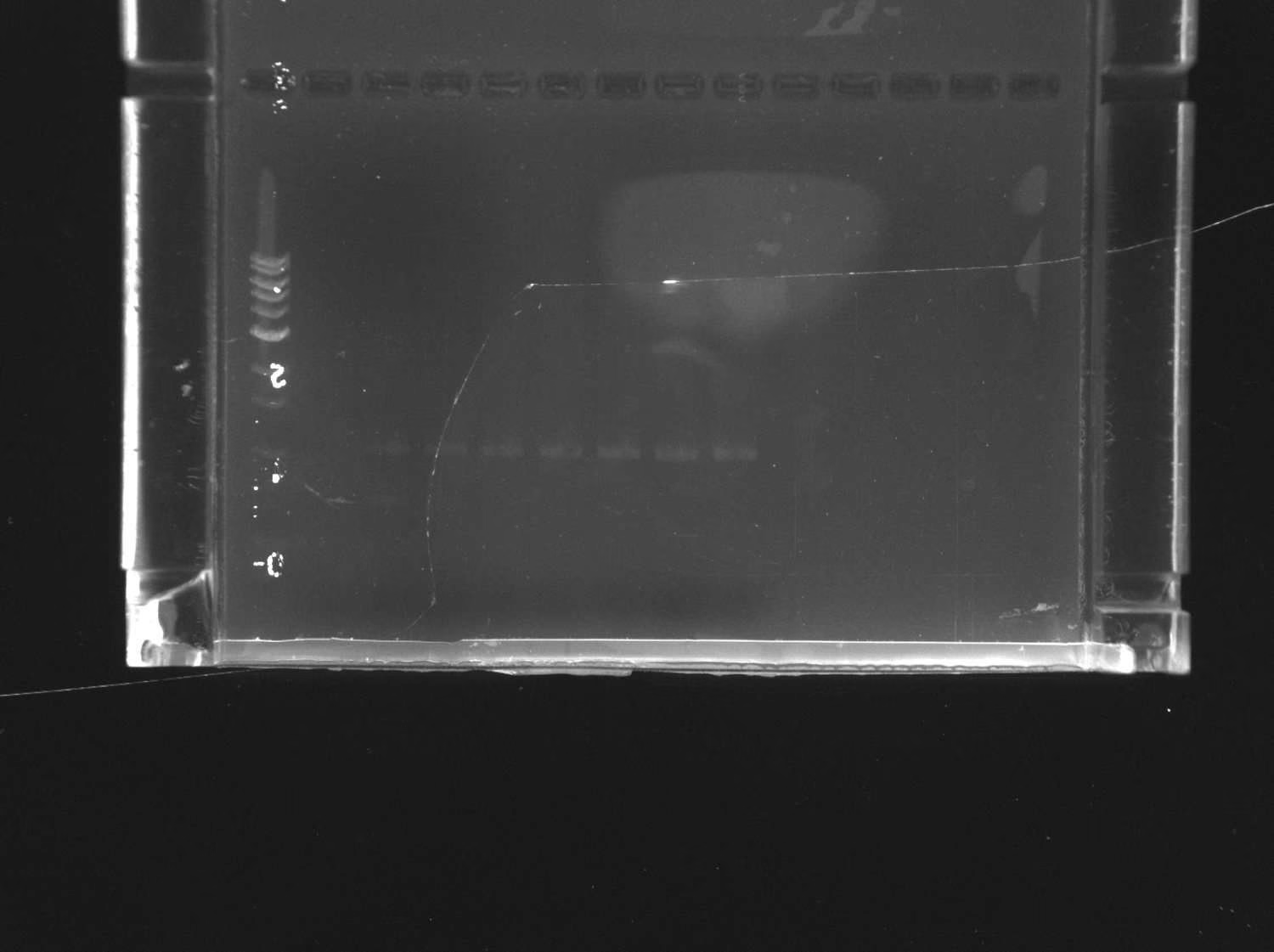
What this shows. Bands fall between 1.0 and 2.0 kb, well below the expected 3 kb product.
Conclusion. The redesigned reaction was amplifying something other than the full plasmid, so this route was abandoned rather than optimised further. Checking product size before committing to a ligation saved a round.
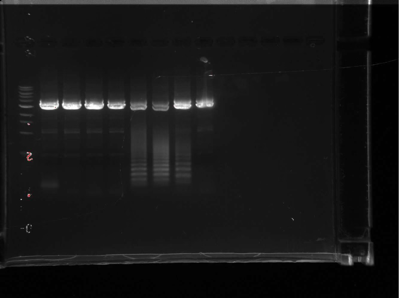
What this shows. Product survives the extended digest, so the amplified DNA is not methylated parental plasmid.
Conclusion. A 30-minute DpnI digest had been leaving enough parental plasmid to dominate the transformation, which explains why sequencing kept returning the starting construct. Overnight digestion became standard.
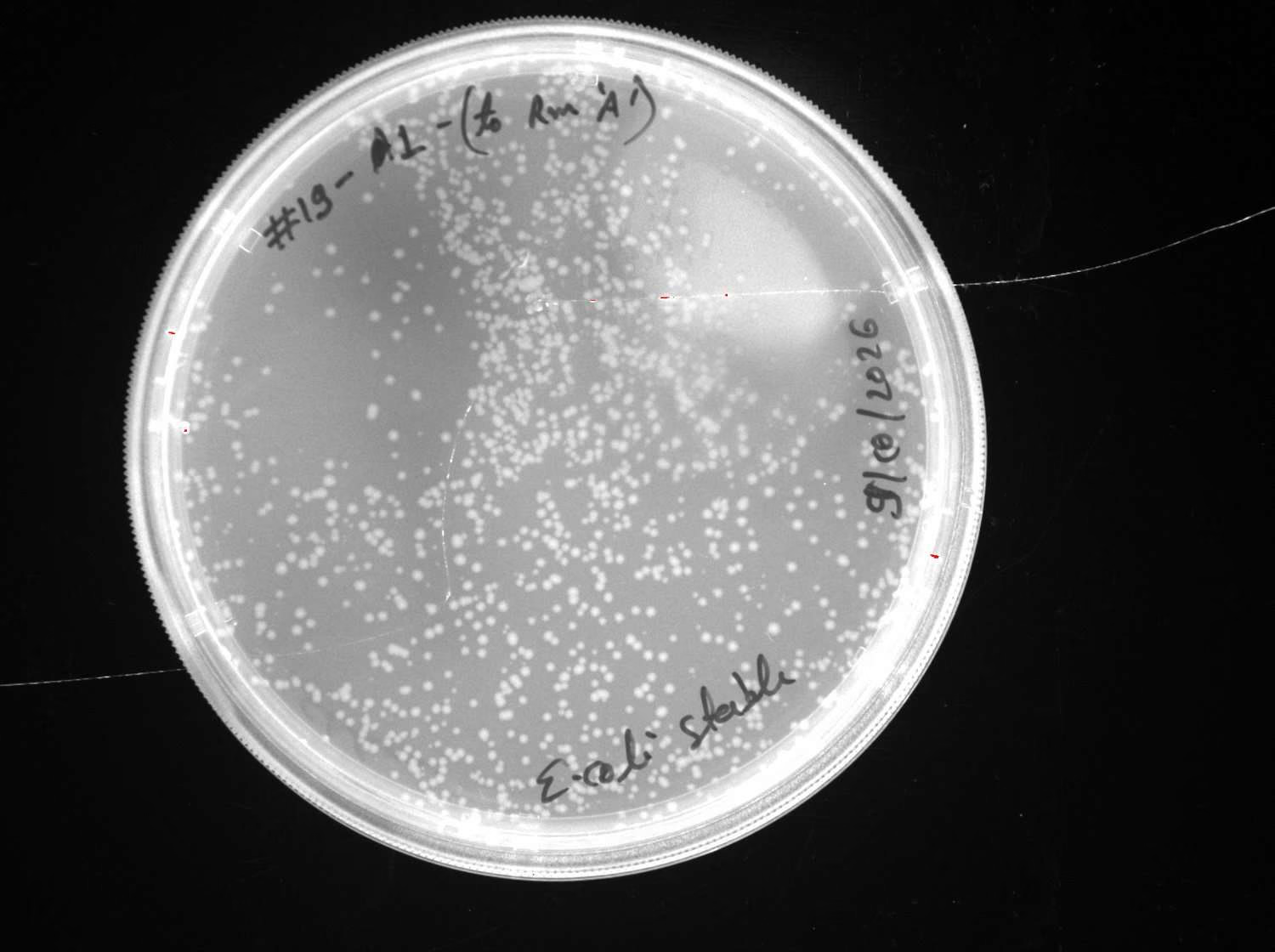
What this shows. Many colonies, markedly more than earlier attempts at the same step.
Conclusion. Colony number alone does not report whether the edit is present, and sequencing of these clones still showed the 'A'. Blunt-end closure inside the cell remained the bottleneck.
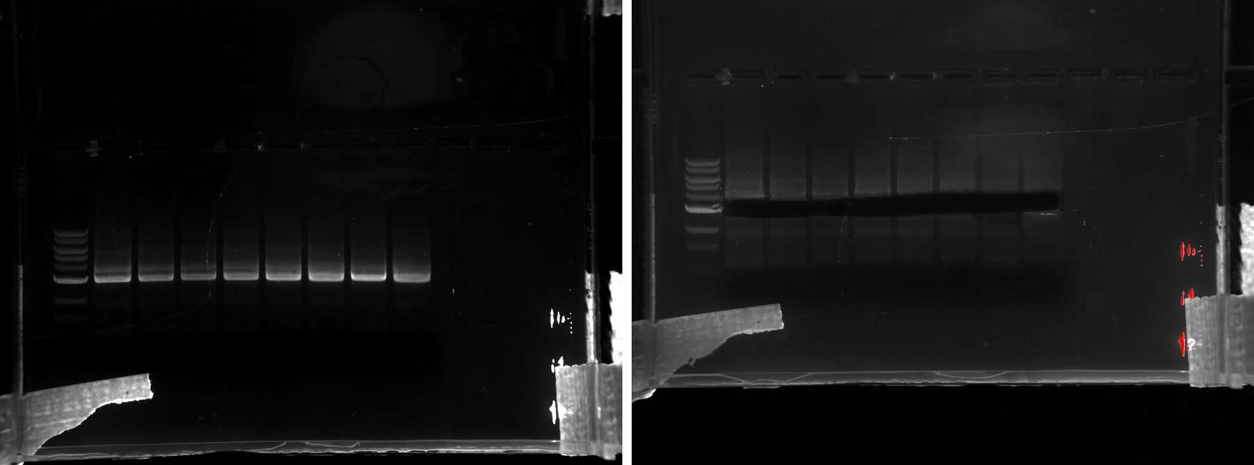
What this shows. A single product at the expected 3 kb, cleanly excised.
Conclusion. This fragment went into the in vitro route: gel extraction, 5′ phosphorylation with T4 PNK, circularisation with T4 DNA ligase, then transformation. PCR products carry no 5′ phosphate, so without the kinase step ligation cannot proceed. That is the step the earlier attempts were missing.
The result that closed the problem
Five colonies (#191 to #195) were submitted for Sanger sequencing, confirming removal of the extra adenine and an intact reading frame in clone #193 while #194 returned unchanged parental plasmid. 6xHis-YebF-ΔTm-FAAH was complete and sequence-verified.Protein expression
Our only quantitative protein result this season. Three constructs were transformed into E. coli BL21(DE3), induced with doxycycline, and separated by SDS-PAGE as uninduced, induced pellet and induced supernatant fractions.
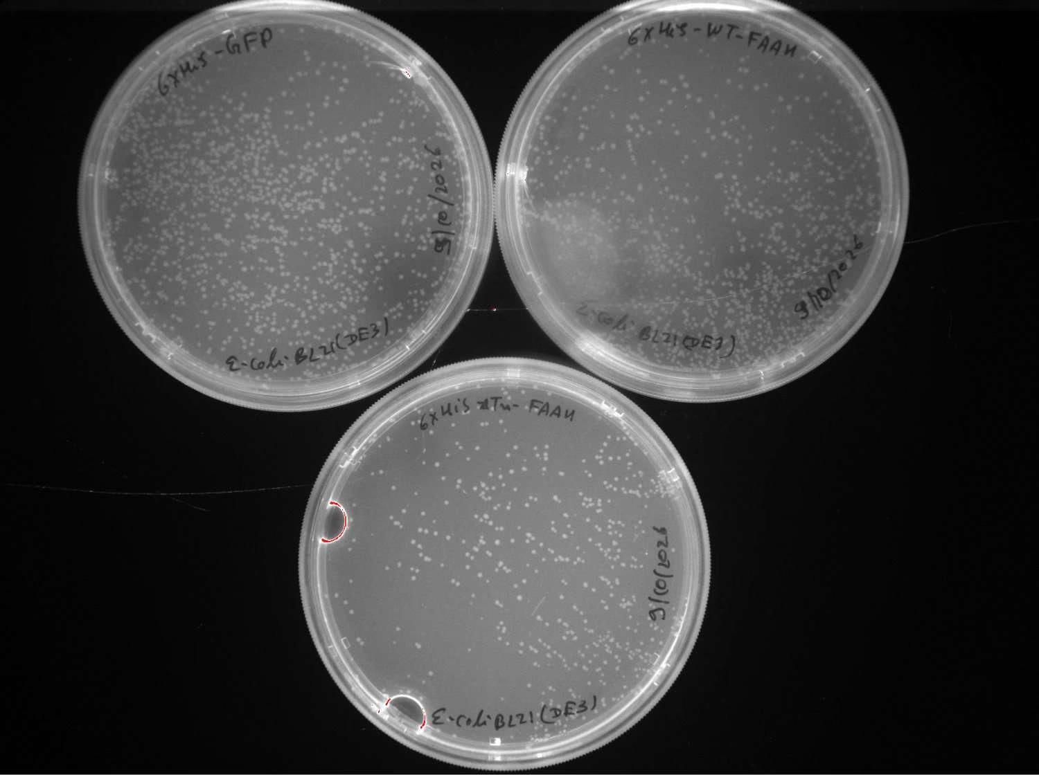
What this shows. All three constructs transformed into the expression strain and recovered colonies.
Conclusion. BL21(DE3) is the correct strain for testing whether a construct expresses, and the wrong strain for the application. These results establish expression, not behaviour in the chassis.
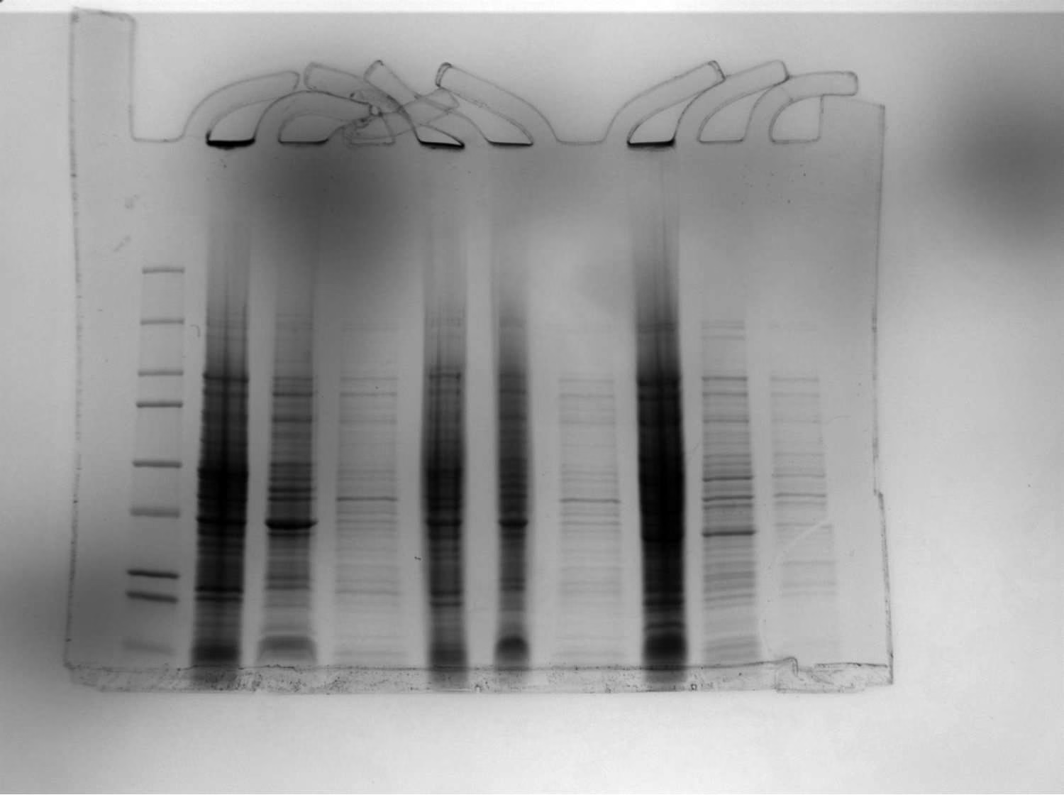
| Construct | Expected mass | Observation |
|---|---|---|
| 6xHis-GFP | 27.7 kDa | Induced band absent from uninduced control |
| 6xHis-FAAH | 44.2 kDa | Induced band absent from uninduced control |
| 6xHis-ΔTm-FAAH | 40.7 kDa | Induced band absent from uninduced control |
What this shows. Each induced construct carries a band that is absent from its own uninduced control, at approximately the expected mass.
Conclusion. All three constructs express on induction. Running pellet and supernatant separately matters for the secretion variants, because which fraction the protein appears in is the result rather than a technical detail.
Limit of this result
An induced band at the right size is evidence of expression, not of identity. Confirming that these bands are our tagged proteins requires the Western blot against the 6xHis tag, which is still outstanding.Transfer into the EcN chassis
The verified secretion construct was moved into E. coli Nissle 1917, the probiotic chassis the project is built around, and banked.
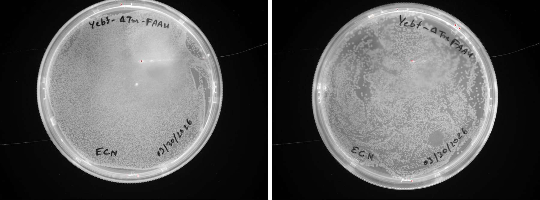
What this shows. EcN takes the verified construct and grows under chloramphenicol selection.
Conclusion. The construct transfers from the cloning strain into the target chassis without further modification, which was not guaranteed: EcN is a robust environmental survivor rather than an attenuated laboratory strain.
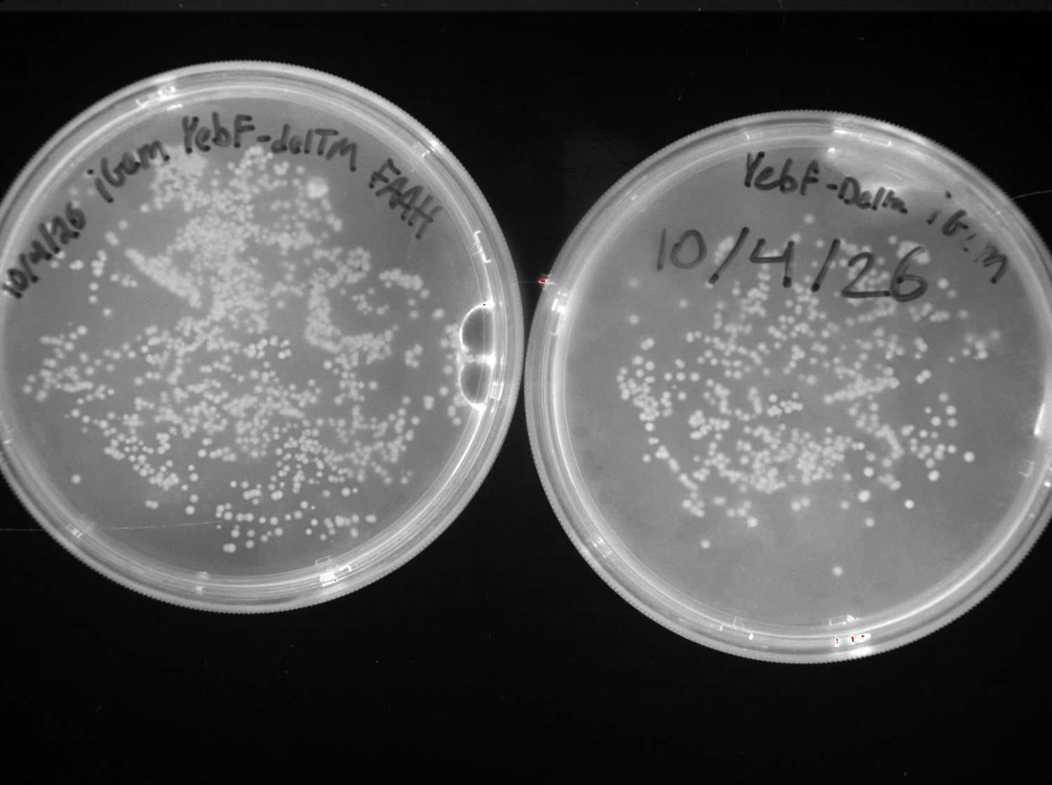
What this shows. Heavy, even growth on both plates from the team transformation run.
Conclusion. This is the first version of the system doing what it was designed to do: a sequence-verified FAAH secretion construct carried by the probiotic chassis. Five glycerol stock tubes were banked, so subsequent work does not depend on repeating the transformation.
Every construct, and where it got to
| Construct | Built | Sequenced | Expressed | In EcN |
|---|---|---|---|---|
| 6xHis-GFP | Yes | Verified | Yes, 27.7 kDa | — |
| 6xHis-WT-FAAH | Yes | Verified | Yes, 44.2 kDa | — |
| 6xHis-ΔTm-FAAH | Yes | Verified | Yes, 40.7 kDa | — |
| 6xHis-YebF-ΔTm-FAAH | Yes | Verified | Not yet | Banked |
| OmpA-ΔTm-FAAH | Primers only | — | — | — |
| OmpX-ΔTm-FAAH | Primers only | — | — | — |
| Catalytic mutant | Not built | — | — | — |
We have not shown that the enzyme is active, that it degrades OEA, or that it changes appetite in anything. The catalytic mutant is the control that makes both activity assays interpretable, so it is the highest-priority item remaining rather than the last.
The dated record behind every figure here is on the Notebook page, the iterations behind them on the Engineering Success page, and the assay designed to test the central claim on the Measurement page.